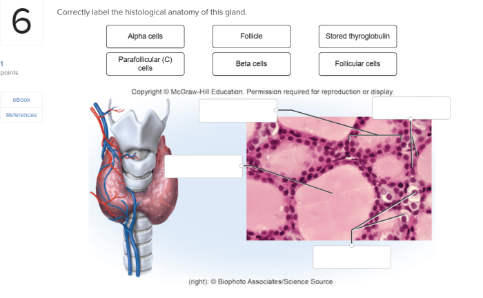
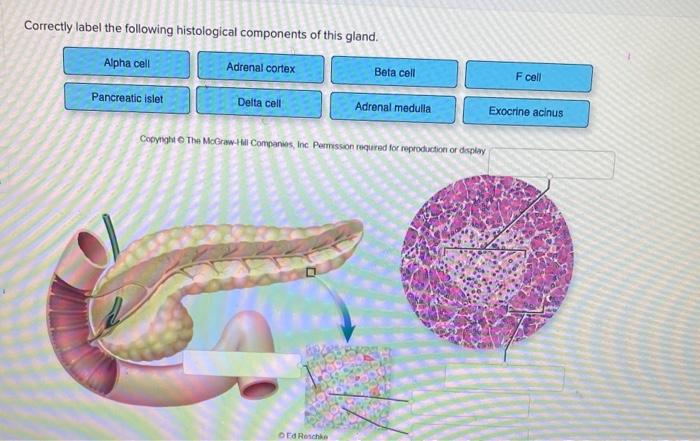
Correctly label the following histological components of this gland. – Correctly labeling the histological components of a gland is essential for accurate diagnosis and treatment. This article provides a comprehensive guide to identifying and labeling the key histological components of a gland, ensuring a thorough understanding of its structure and function.
Histological Components of the Gland: Correctly Label The Following Histological Components Of This Gland.

The gland is composed of several histological components, each with a specific structure and function. These components include:
-
-*Epithelial cells
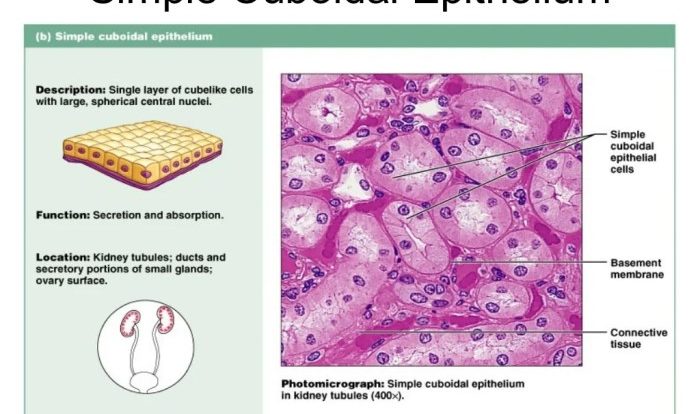
The epithelial cells line the ducts and acini of the gland. They are responsible for secreting the gland’s products.
-*Myoepithelial cells
The myoepithelial cells are located between the epithelial cells and the basement membrane. They are responsible for contracting the gland and expelling its products.
-*Basement membrane
The basement membrane is a thin layer of connective tissue that separates the epithelial cells from the underlying stroma.
-*Stroma
The stroma is the connective tissue that surrounds the epithelial cells and myoepithelial cells. It provides support and nourishment to the gland.
Labeling the Histological Components

To correctly label the histological components of the gland, follow these steps:
- Identify the epithelial cells. They are the cells that line the ducts and acini of the gland.
- Identify the myoepithelial cells. They are the cells that are located between the epithelial cells and the basement membrane.
- Identify the basement membrane. It is the thin layer of connective tissue that separates the epithelial cells from the underlying stroma.
- Identify the stroma. It is the connective tissue that surrounds the epithelial cells and myoepithelial cells.
Importance of Correct Labeling

Correctly labeling the histological components of the gland is important because it allows pathologists to accurately diagnose and treat diseases of the gland. For example, if a pathologist incorrectly labels the epithelial cells as myoepithelial cells, they may misdiagnose a cancer of the gland as a benign condition.
Applications of Correct Labeling

Correctly labeling the histological components of the gland has a number of applications in research, diagnosis, and treatment. For example, correctly labeling the histological components of the gland can be used to:
- Diagnose diseases of the gland
- Develop new treatments for diseases of the gland
- Study the normal function of the gland
Question Bank
What are the key histological components of a gland?
The key histological components of a gland include the secretory cells, ducts, and supporting tissues.
Why is it important to correctly label the histological components of a gland?
Correctly labeling the histological components of a gland is important for accurate diagnosis and treatment. Incorrect labeling can lead to errors in diagnosis and treatment, potentially affecting patient outcomes.
